The Cancer Systems Microscopy Team
We have multiple Positions Open - scroll down to see student and staff positions available
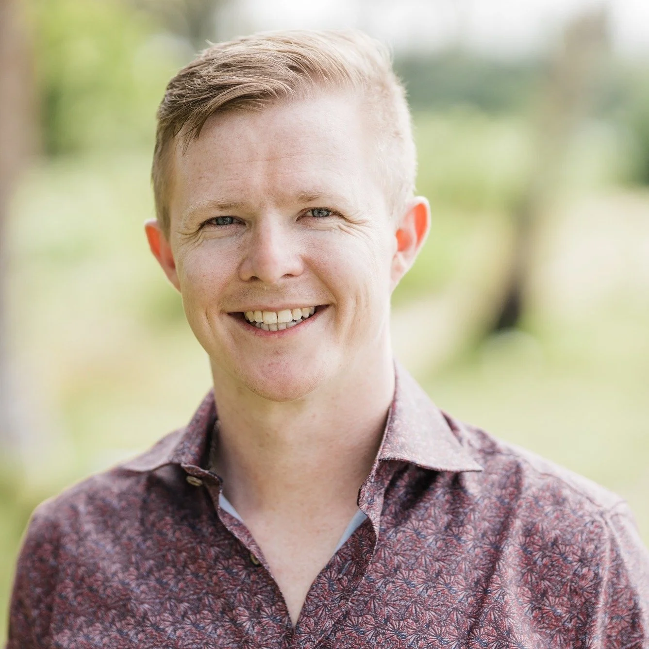
John lock, group leader
John is passionate about both fundamental and translational cancer research. He tackles these topics primarily through the application of imaging-based systems biology techniques; collectively termed ‘Systems Microscopy’. This reflects John’s role as an early pioneer in quantitative microscopic imaging and analysis of live and fixed single cells - using these tools to interrogate complex molecular systems, emergent cell behaviours and heterogeneity across these scales in time and space. John has driven such research into many aspects of cancer cell biology including cell phenotype / morphology, cell-cell and cell-matrix adhesion, cell migration and polarity, cell division, protein trafficking and various molecular signalling systems.
An early adopter of quantitative live and fixed single cell imaging during his research into protein trafficking and sorting mechanisms (PhD, Institute for Molecular Bioscience, University of Queensland), John next went on to establish a multidisciplinary Systems Microscopy team (cell biologists, biophysicists, statisticians) exploring cancer cell adhesion and migration biology (Postdoc / Assistant Professor, Karolinska Institute, Sweden). This team helped to found a large-scale EU-funded Network of Excellence for Systems Microscopy dedicated to advancing all aspects of this new research modality (high-throughput experimentation, automated microscopy, image quantification, statistical analysis, machine vision). Focused on the biology of cell adhesion, migration and mitosis, this network included leaders in advanced approaches including functional genomics, high-throughput drug screening and imaging-based precision medicine. Over 5 years, this network delivered key insights - both specific and systemic - into fundamental as well as translational aspects of cancer cell biology.
Since returning to Australia and establishing the Cancer Systems Microscopy Lab (University of New South Wales, Sydney), John has continued his commitment to driving innovative advances across the multidisciplinary Systems Microscopy pipeline, whilst increasing his biological focus on translational (clinical) applications of his cancer cell biology research. Key outcomes already include playing a leading role in formation of the Ramaciotti Systems Microscopy Unit at UNSW - dedicated to imaging-based systems biology, especially Proteomic Microscopy. John and his collaborators have also received Australian Research Council (ARC) and National Health & Medical Research Council (NHMRC) grants for projects including statistical analysis of signaling pathway activities and diagnostic imaging of circulating tumour cells, as well as UNSW grants funding microscopes, robotics hardware and data visualisation software development. John is also actively engaged in steps to commercialise his recent advances in drug discovery and data visualisation.
With a growing research team within the Faculty of Medicine at UNSW and a recently formalised affiliation with the Ingham Institute for Applied Medical Research (Liverpool, Sydney), John is now excited to focus on research projects with the maximum potential to improve cancer patient outcomes through development of precision diagnostics, accelerated drug discovery and a deeper fundamental understanding of cancer cell systems biology.
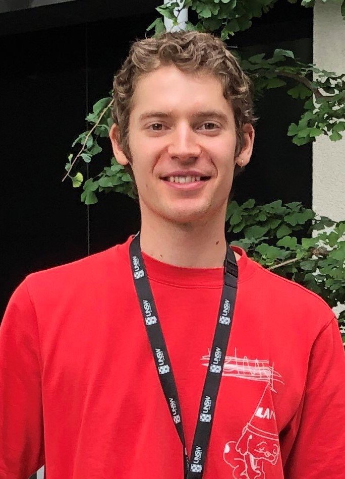
Daniel Neumann – Post-Doctoral Fellow
Daniel is driven to understand how non-genetic mechanisms of gene regulation contribute to the diversity of cancer cell states and behaviours. His hope is that the study of these mechanisms will lead to the development of novel therapeutics that will prevent the most troubling aspects of cancer progression, particularly metastasis and drug resistance.
Daniel studied a Bachelor of Medical Science through the University of South Australia and the Australian National University in a co-badged program. His interest in the world of mRNA regulation led him to pursue a PhD in the Centre for Cancer Biology under the supervision of Philip Gregory and Greg Goodall. His work was focussed on how the isoforms of RNA-binding protein QKI cooperate to regulate epithelial cell plasticity, and how the nuclear isoform, QKI-5, regulates alternative splicing and polyadenylation.
As a Post-Doctoral Fellow at the Cancer Systems Microscopy Lab, Daniel is continuing his study of cancer cell plasticity, focussing on the regulatory networks governed by the transcription factor, ZEB1. Specifically, he will use live cell fluorescence microscopy to map expression and localization of ZEB1 to the molecular changes and shifts in cell states and behaviours that are observable in the cancer plasticity landscape.
When he is not at the lab, Daniel enjoys playing guitar and piano. He also likes to go on nature walks and explore Sydney with his partner.
Tim Mann - Post-doctoral fellow
Tim is passionate about translational science – converting scientific discoveries in the lab into real world impacts.
Tim completed his PhD in 2022 with Western Sydney University at the Ingham Institute for Applied Medical Research (Liverpool), investigating interactions of pro-inflammatory protein hGIIA in prostate cancer, as well as the function of its inhibitor c2, developed by supervisor A/Prof. Kieran Scott. Using a range of live cell and high-resolution imaging techniques, Tim discovered novel interactions of hGIIA which contribute to its ability to promote prostate cancer growth, as well as the mechanisms via which c2 inhibits this. Following this, he continued this research in a post-doctoral position with Australian start-up biotechnology company Filamon, which has led to the filing of multiple patents, and the development of an exciting new class of cancer therapeutics derived from c2.
This year Tim has started in the Cancer Systems Microscopy lab here at UNSW as a post-doctoral researcher developing methods to guide cancer therapy. Now with the vast array of treatment options available, the critical step is getting the right treatment to the right patient, as every cancer is different and constantly changing. Leaning on his previous expertise in confocal imaging and the development and investigation of novel cancer therapeutics, Tim will be developing multiplexing protocols for use in cancer patients’ liquid biopsies. Specifically, this will entail collecting blood samples from cancer patients undergoing treatment, extracting the cancer cells present in these samples and applying a systems microscopy approach (including imaging of many different proteins) and quantitative analysis to identify novel biomarkers to better predict treatment efficacy and resistance.
Outside of the lab, Tim enjoys spending time with his wife and dog, as well as gaming, kayaking and watching the footy - up the Giants!
gloria (ye) zheng - research assistant
Gloria spent two years (2018-2020) working to understand key signaling pathways in colorectal cancer and colon organoid cultures, as part of her studies for a Masters of Biomedical Science at the University of Melbourne (Burgess Laboratory, Walter and Eliza Hall Institute of Medical Research). Upon completing her Masters, she worked as a research assistant in the Burgess Laboratory using quantitative imaging to monitor the anti-cancer effects of a new drug impacting mitosis and apoptosis in cancer cell lines.
She has great enthusiasm to continue expanding her knowledge and understanding of microscopy, particularly in cell biology and cancer research, as well as having a real passion to undertake a PhD in the near future.
Having studied violin for 6 years, Gloria likes listening to music - especially classical music. She also enjoys reviewing great movies, photographing nature and experiencing different cultures, reading books and cooking.
Felix Kohane, PhD Student - UNSW School of medical sciences
Felix is inspired by the possibilities of using innovative multi-disciplinary strategies for understanding complex biological systems and developing precision diagnostic tools for cancer patients. He developed an interest in computational biology and artificial intelligence during his Honours year at the Garvan Institute of Medical Research (2020), where he examined the mechanisms behind non-genetic cell-state changes contributing to the complex and heterogenous nature of triple-negative breast cancers. His research employed high-throughput image-based profiling of single-cells combined with an application of dimension reduction and machine learning methods to model the system and extract meaningful relationships between cell subpopulations.
In his PhD, Felix will extend his knowledge and application of computational and AI-based analytical tools, including developing deep learning models for cellular analysis and applying novel single-cell RNA-seq algorithms to proteomic image data. He is particularly interested in expanding these tools into a clinical setting through developing the lab’s capacity for proteomic microscopy, allowing for biomarker characterisation in patient-derived circulating tumour cells.
Outside of the lab, Felix’s latest passion is AI-generated art and design, and his interests include architecture, fashion and art history. He enjoys seeing good films, music of all kinds and attempting to cook.
Andrew Gunawan, PhD Student - UNSW Engineering & unsw medicine
Andrew completed his UNSW undergraduate degree in Computer Science and then his Honours year in the Wilkins lab (UNSW Faculty of Science, BABS), studying protein structures and post-translational modification-induced conformational changes. By conducting a bioinformatics analysis of mass spectrometer-identified chemical crosslinks in proteins, he has been able to fuse his passion for computer science and biology. In his PhD, Andrew is keen to bring computer vision to cell microscopy, aiming to push the boundaries in computer vision, but also to contribute to our understanding of life.
In his spare time, Andrew enjoys talking to his koi fish and pot plants (though they rarely talk back). He has also been known to lose a lot at table tennis but will accept any challengers wanting a match.
chantelle johnstone, Honours Student - UNSW School of biomedical sciences
Chantelle is keen to play a part in the Cancer Systems Microscopy Lab's contributions to emerging research developing precision diagnostics for cancer. She has recently completed a Bachelor of Medical Science (Pathology) with the University of New South Wales. Her favourite topics during this degree were cancer pathology and learning about the visualisation of disease using emerging imaging techniques.
As an Honours student, Chantelle will be refining imaging-based analysis of signalling pathways in non-small cell lung cancer. Plans to translate these methods to blood sample-based circulating tumour cell analysis aims to better match patient heterogeneity to targeted treatments. The use of immunofluorescence imaging and computational analysis of high dimensional data at the CSM lab is of great interest, and Chantelle is keen to explore this area in her year of research.
In her spare time, Chantelle enjoys exploring new places with friends, nature walks, bouldering and music.
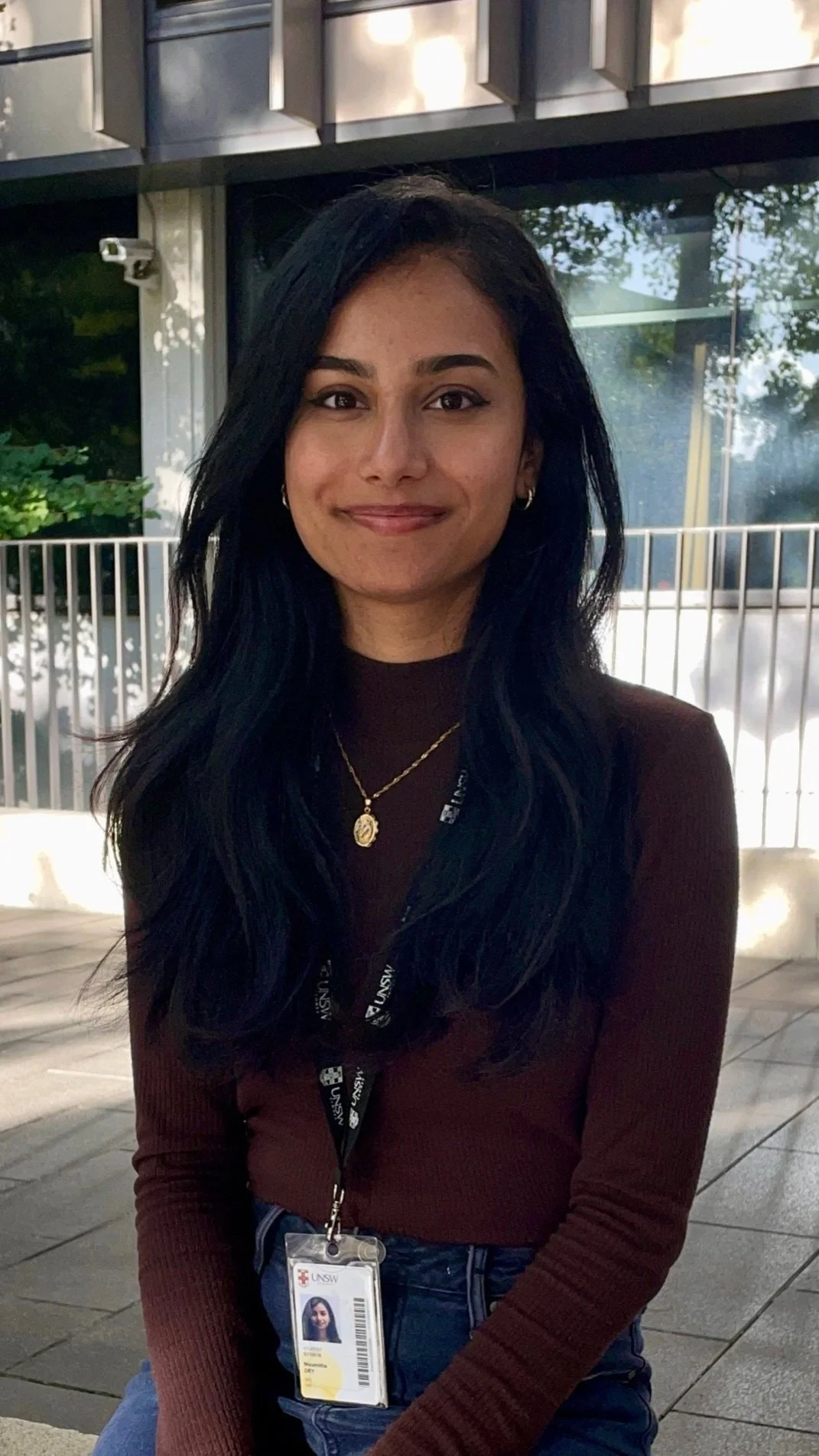
Moumitha dey, Honours student - unsw school of biomedical sciences
Moumitha is interested in extending her pharmacology knowledge and skills through the multi-disciplinary approaches she will engage in over her Honours year with the CSM lab. She is enthusiastic about the emerging applications of computational biology and the role of AI and machine learning in the cancer research field, particularly in tackling cancer heterogeneity and drug resistance, along with other precision medicine tools.
As part of her undergraduate Advanced Science degree at UNSW (Pharmacology), Moumitha will be venturing into the field of pathology, picking up new skills in tissue culture, immunofluorescence, microscopy, multiplexing and computational analysis. Empowered by these skills, she aims to contribute to the development of a novel and data-driven diagnostic tool able to guide treatment of prostate cancer patients through comprehensive analysis of their molecular characteristics.
Outside of the lab, Moumitha holds a black belt in Taekwondo but mostly sticks to her sewing projects, listening to podcasts, and catching sunsets at Sydney’s beaches even though she can’t swim.
Open Positions - 2 PhD Candidates
We are currently seeking 2 PhD candidates who are passionate about contributing to translational cancer research and have training / experience in (cancer) cell biology, fluorescence microscopy, quantitative image analysis, and/or multivariate statistics / machine learning / deep learning. Though you may not currently have expertise in all of these areas, creativity, curiosity and a drive to learn across these disciplines will be your greatest assets and will ensure you have highly sought after skills and diverse professional options upon completion of your PhD studies.
You will be engaged as part of a collaborative team in a fully funded, world-first research project applying Systems Microscopy techniques to achieve precision diagnosis of cancer-drivers in advanced cancer patients. Specifically, you will perform “Proteomic Microscopy” (see our methods page) on circulating tumour cells (CTCs) isolated from cancer patient liquid biopsy blood samples. Using image analysis, advanced statistical tools, machine learning / deep learning and novel VR-based data visualization tools, you will help to define biomarker signatures that will enable next generation precision cancer medicine through the selection and development of pathway-specific therapeutics.
In parallel to this core collaborative project, you will have the opportunity to develop your independence by driving fundamental research aimed at understanding signalling mechanisms in related cancer cell models. This will ensure diversification of your research outcomes and provides additional avenues for you to develop your research capacity.
With excellent communication skills (written and spoken English) and commitment to both team work and independent research / learning, you might be well-suited to this project. So feel free to contact us through our contacts page, or email John directly at john.lock@unsw.edu.au.
Open Position - Honours Student
We have several exciting research projects suitable for Honours students, ranging across our multidisciplinary Systems Microscopy pipeline. Depending on your training background and goals, you could contribute to projects focused on or combining: cancer (especially prostate or lung) cell signalling biology; automation of imaging and liquid handling; image analysis including machine vision and deep learning; statistical analyses including manifold embedding and feature selection; information theory approaches and causal modelling; data visualisation including developing / testing interactive VR-AI feedback methods. Get in contact through our contacts page, or email John directly at john.lock@unsw.edu.au.


